Opening Hours: Mon - Fri 8.00 - 16.30

3MV Tandetron

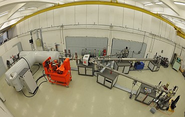
The 3 MV Tandetron™ accelerator system has been installed and commissioned in 2012 at the "Horia Hulubei" National Institute for Physics and Nuclear Engineering - IFIN-HH, Măgurele, Romania [1]. The main purpose of this machine is to strengthen applied nuclear physics research ongoing in our institute for more than four decades. The accelerator system was developed by High Voltage Engineering Europa B.V. (HVE) and comprises three high energy beam lines. The first beam line is dedicated to ion beam analysis (IBA) techniques: Rutherford Backscattering Spectrometry - RBS, Nuclear Reaction Analysis - NRA, Particle Induced X-ray and γ-ray Emission - PIXE and PIGE and micro-beam experiments - μ-PIXE. The second beam line is dedicated to high energy ion implantation experiments and the third beam line was designed mainly for nuclear cross-sections measurements used in nuclear astrophysics. A unique feature, the first time in operation at an accelerator facility is the Na charge exchange canal (CEC), which is used to obtain high intensity alpha beams of at least 3 μA. The system in Bucharest is based on a 3 MV Tandetron™ that uses an all-solid-state parallel-fed Cockcroft–Walton power supply for generating high-voltage [2] and an ion beam injector comprising two negative ion sources.

[1] I Burducea et al., Nuclear Instruments and Methods in Physics Research B, 2015
[2] C Podaru et al., Nuclear Instruments and Methods in Physics Research B, 2013


3 MV Tandetron webpage
secretar.dfnanipne.ro

The first beam line is equipped with a multipurpose Ion Beam Analysis (IBA) chamber for: Rutherford backscattering spectrometry—RBS, foil-Elastic Recoil Detection Analysis—ERDA, particle-induced X-ray and gamma-ray emission—PIXE and PIGE and micro-beam experiments—micro-PIXE. The HVE data acquisition software records spectra from several detectors simultaneously, capturing RBS, ERDA, PIXE and PIGE signals. In other words, total IBA analysis can be performed. It is worth noting that IBA chamber is also equipped with three-axis (phi, theta and tilt) computer-controlled goniometer that enables quantification of the disorder in ion-irradiated single crystals via Rutherford backscattering spectrometry in channeling configuration (RBS/C) measurements. Lattice site location of impurities can be also determined from the analysis of RBS/C spectra. It is also worth nothing that the ion track diameter can be also deduced from cross-section measurements resulting from the analysis of RBS/C data.

Top-view of IBA reaction chamber with detectors (a) and sample holder goniometer (b)

Responsible: Dr. Ion Burducea
bionnipne.ro


The external beam setup was designed and implemented to serve for radiobiology studies, but also to extend the artefacts range measured for archaeometry. Self-supported Si3N4 membranes proved to be of paramount interest during the last three decades, mainly due to their mechanical strength, optical properties and also for various beams (X-ray, electrons and ions) scattering properties.. Disposing of a thin membrane that withstands 10 orders of magnitude pressure difference, while minimally altering the ion beam properties, represents a huge asset for MeV-range accelerators. Thus, one may perform in-air experiments with ion beams on liquid samples or analyze huge objects that in most cases do not fit in a common vacuum chamber. One of the main challenges for an external setup resides in performing a precise beam characterization, which was achieved in our case either via live monitoring with solid-state silicon detector installed in vacuum, in RBS geometry or by using a Markus ionization chamber, plastic track detectors and radiochromic films for in-air dosimetry.

External beam setup 3D schematics

Responsible: Dr. Mihai Straticiuc
mstratnipne.ro

The second beam line is equipped with a dedicated chamber for high-energy ion irradiation and implantation experiments. A four corner Faraday cup (4 IIB FC) assembly is positioned before the implantation chamber to measure the implanted dose. These 4 IIB FC are also used to define the scanned area (irradiation area) which is a square with a fixed side of 125 mm. Since the position of these 4 IIB FC cannot be changed, high irradiation fluences (e.g., 150 ions/nm2) cannot be achieved within a reasonable time. Thus, a new set of 4 IIB FC that defines a square with much smaller side (29 mm) was developed in-house. This ongoing upgrade enables a high dose within reasonable time, which is relevant in the field of nuclear materials research for fission/fusion reactor systems or to produce the nanoparticles by ion beam synthesis technique. Since during the irradiation stage it is crucial to monitor irradiation temperature, the installation of two thermocouples has been made: one monitors the temperature on the backside of the sample holder and the second one determines the temperature on the sample surface. The direct comparison between data collected from these two thermocouples and infrared camera allows the precise determination of the sample temperatures.

Examples of ionic species* that can be accelerated at 3 MV Tandetron: https://dfna.nipne.ro/3MV_Tandetron_EN.php

Irradiation sample holder: liquid nitrogen cooled (left) and heated at 800o C (right)

Responsible: Dr. Gihan Velisa
gihan.velisanipne.ro


The third beam line is a multipurpose one and can be used for instance for nuclear cross section measurements (CSM) used in nuclear astrophysics. An external-beam nozzle for ion irradiation of biological samples and external beam PIXE for large samples analysis in air is connected to the exit port of the IBA chamber. This upgrade was done in-house.

Responsible: Dr. Alexandra Spiridon
alexandra.spiridonnipne.ro

IFIN-HH / ELI-NP User Office

Contact Us

No. 30, Reactorului Street, Magurele, Ilfov, Romania (077125)
+40(0)21.404.23.01